Chronic hepatitis E mostly occurs in organ transplant recipients and can lead to rapid liver fibrosis and cirrhosis. Previous studies found that the development of chronic hepatitis E virus (HEV) infection is linked to the type of immunosuppressant used. Animal models are crucial for the study of pathogenesis of chronic hepatitis E. A research group led by Dr. Lin Wang in Peking University, Beijing, China previously established a stable chronic HEV infection rabbit model using cyclosporine A (CsA), a calcineurin inhibitor (CNI)-based immunosuppressant. However, the immunosuppression strategy and timing can be further optimized, and how different types of immunosuppressants affect the establishment of chronic HEV infection in this model is still unknown.
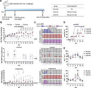
Recently, Dr. Lin Wang showed that chronic HEV infection can be established in 100% of rabbits when CsA treatment was started at HEV challenge or even 4 weeks after (Figure 1).

Fig 1 Chronic HEV infection in rabbits when starting CsA treatment at different timepoints of infection. (A) Experimental scheme. CsA treatment started at the same time of HEV-3ra challenge (CsA-0w group) or 4 weeks after HEV-3ra challenge (CsA-4w group). Rabbits not treated with CsA were also set as an acute infection control (no-CsA group). (B) Quantification of fecal HEV RNA, serum HEV RNA, and serum HEV antigen over time after inoculation. (C) Number of rabbits with HEV fecal shedding, viremia, or antigenemia over time after inoculation. (D) Weekly anti-HEV positive rates. (E) Serum ALT and AST levels. Error bars indicate mean ± SEM. S/CO values of >1 were considered positive. n = 4 per group, *P < 0.05, **P < 0.01, ***P < 0.001. ALT, alanine aminotransferase; AST, aspartate aminotransferase; LOQ, limit of quantification; S/CO, signal-to-cutoff.
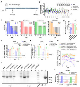
Tacrolimus or prednisolone treatment alone also contributed to chronic HEV infection, resulting in 100% and 77.8% chronicity rates, respectively, while mycophenolate mofetil (MMF) only led to a 28.6% chronicity rate (Figure 2).

Fig 2 Chronic HEV infection in rabbits treated with MMF, tacrolimus, or prednisolone alone. (A) Experimental scheme. The MMF group (n = 11), the tacrolimus group (n = 11), and the prednisolone group (n = 13) were inoculated with HEV-3ra and treated with one kind of immunosuppressant alone, respectively. The untreated group was only inoculated with HEV-3ra (n = 10). The negative group (n = 8) was only mock inoculated. Four rabbits from each group were chosen to be sacrificed at 4 wpi and the remaining were sacrificed at 13 wpi. (B and C) Viral load in feces from each rabbit and numbers of rabbits with viral fecal shedding over time after inoculation in HEV-infected groups. (D) Duration of viral fecal shedding in HEV-infected groups. (E–G) HEV RNA determined by RT-qPCR (E) and negative-stranded HEV RNA detected by RT-qPCR (F). Nested PCR (G) in livers from rabbits sacrificed at 4 or 13 wpi in the HEV-infected groups. (H) Viral load in feces from the 10 naïve rabbits challenged with inoculums 1–5. (I) Anti-HEV seroconversion of the MMF group, the tacrolimus group, the prednisolone group and the untreated group at 0, 4, 8 and 13 wpi. HEV, hepatitis E virus; wpi, week-post-inoculation; LOQ, limit of quantification; *P < 0.05, **P < 0.01, ***P < 0.001. N, negative; NC, negative control; P, positive; PC, positive control; RT-qPCR, real-time quantitative PCR.
Chronic HEV infection was accompanied with a persistent activation of innate immune response evidenced by transcriptome analysis. The suppressed adaptive immune response evidenced by low expression of genes related to cytotoxicity (like perforin and FasL) and low anti-HEV seroconversion rates may play important roles in causing chronic HEV infection. By analyzing HEV antigen concentrations with different infection outcomes, we also found that HEV antigen levels could indicate chronic HEV infection development. This study optimized the immunosuppression strategies for establishing chronic HEV infection in rabbits and highlighted the potential association between the development of chronic HEV infection and immunosuppressants.
IMPORTANCE
Organ transplant recipients are at high risk of chronic hepatitis E and generally receive a CNI-based immunosuppression regimen containing CNI (tacrolimus or CsA), MMF, and/or corticosteroids. Previously, we established stable chronic HEV infection in a rabbit model by using CsA before HEV challenge. In this study, Dr. Lin Wang further optimized the immunosuppression strategies for establishing chronic HEV infection in rabbits. Chronic HEV infection can also be established when CsA treatment was started at the same time or even 4 weeks after HEV challenge, clearly indicating the risk of progression to chronic infection under these circumstances and the necessity of HEV screening for both the recipient and the donor preoperatively. CsA, tacrolimus, or prednisolone instead of MMF significantly contributed to chronic HEV infection. HEV antigen in acute infection phase indicates the development of chronic infection. Their results have important implications for understanding the potential association between chronic HEV infection and immunosuppressants.
Read the full article (J Virol. 2024 Jun 20:e0084624): DOI: 10.1128/jvi.00846-24

